Associates for Surgical Care
Vero Beach, Fla.
General anesthesia was once provided using a face mask and a chin lift held by my left hand. My right hand would adjust the “APL” valve and rest upon the full reservoir bag. The living, breathing patient could be sensed and assisted beneath my fingers as I gently rode the bag to the rhythmic “to and fro” of respiration by stethoscope. The patient and practitioner functioned as one.
The Fundamental Challenge
The upper airway obstruction of general anesthesia is comparable to that of obstructive sleep apnea (OSA).1 General anesthesia, produced by inhalational or IV agents, or their “balanced,” “multimodal” combinations, will cause collapse of the soft tissues of the upper airway.
As happens in OSA, nasal positive airway pressure can open the airway to enable spontaneous respirations.2 Anesthesiologists, in their preoccupation with invasive devices to secure the airway, have been distracted from the usefulness of noninvasive devices to avoid the apnea of induction and the other hazards created by conventional airway management.3,4
I have been slow to appreciate the usefulness of nasal positive pressure even though it has been on and off my mind for half of my 51 years of anesthesia practice. Let me give a brief history.
Years ago, I joined a very progressive plastic surgeon, and the anesthesia he had become accustomed to was monitored anesthesia care (MAC) that, in effect, frequently became total IV anesthesia (TIVA). Oxygen was given by nasal cannula with pulse oximetry as the respiratory monitor. With the coming of laser resurfacing, the fire hazard became dramatic, so solutions evolved.
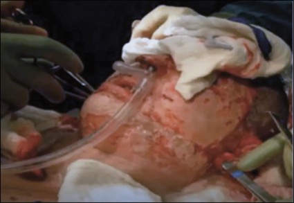
At first, a nasopharyngeal airway was connected by a length of tubing to the anesthesia circuit.5 Then, a fire-resistant metal device was developed which could be inserted “awake” into the nasal vestibule to deliver CPAP from the combination of the anesthesia machine with a CPAP machine. A circuit stethoscope replaced the suprasternal stethoscope to detect upper airway obstruction and facilitate CPAP adjustment (Figure 1).6 The “pop-off” of excess gases was usually through the mouth, but occasionally the leakage was so minimal as to allow rebreathing through the circuit with the detection of end-tidal carbon dioxide (CO2).
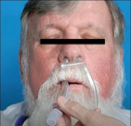
The patients did well, and eventually I came to appreciate that my MAC anesthetics were mostly TIVA anesthetics. The side effects that I once thought were avoided with light sedation were really avoided by maintaining an unobstructed airway with nasal CPAP. Judiciously applied, the required airway pressures did not “blow up” the stomach or create regurgitation or secretion problems, even in procedures lasting several hours. In fact, I began to use nasal CPAP to deeply extubate my general anesthesia patients and to transport them to the recovery room (Figure 2).
Years passed, my practice setting changed, and then a threatened shortage of propofol led me to try mask inductions in adults as I did in children. Sevoflurane worked easier and quicker! Then, it occurred to me that modifying a commercially available nasal CPAP mask to make it compatible with an anesthesia circuit offered the advantage of uninterrupted oxygenation and depth of anesthesia all the way from induction through placement of an invasive airway. With the vent holes of the mask closed by silicone sealant, sufficient positive pressures could be generated within the gas flow capacity of the anesthesia machine. Now, I could coach a patient to unresponsiveness within two to four breaths of ... “In through the nose! … Hold it! One thousand one; one thousand two … Out through the mouth!” I used 8% sevoflurane in 100% oxygen, and in short order, the mouth could be opened for the insertion of a laryngeal mask airway or a Macintosh laryngoscope.
For intubation, with visualization of the vocal cords, topical anesthesia could first be sprayed to cause a mild cough and a short-lived laryngospasm. Spontaneous respirations returned immediately, and an endotracheal tube could be placed without further reaction or interruption of breathing.
If a surprisingly difficult airway should develop, then oxygenation, depth of anesthesia and spontaneous respirations could be maintained indefinitely while working out a solution. Meanwhile, gas flows are within the capacity of the anesthesia machine to produce pressures sufficient to open the airway and to facilitate manually assisted respirations. With sevoflurane, the worrisome reflexes of swallowing, secretions and vomiting are suppressed early upon induction while the airway-protective reflex of laryngospasm is preserved down to surgical levels of anesthesia. The need to scavenge the waste anesthesia gases is met by inserting a “wall suction’’ into an overlying face mask.
{RELATED-HORIZONTAL}Summary of the Solution
In my opinion, for every elective general anesthetic, nasal positive pressure provides the safest, most useful airway management during both induction and emergence. The maintenance stage posed the remaining question for me: Can the airway be secure throughout a prolonged volatile anesthetic? And my answer is why not! I had used the technique safely for several years through hundreds of TIVAs—obstruction from a volatile agent is no different than that caused by an IV agent. If anything, by Guedel’s observations,7 sevoflurane is quick to suppress the annoying reflexes while preserving the protective reflex of laryngospasm.
Recently, I have used nasal positive pressure from start to finish in consecutive patients without mitigation for obesity, bushy facial hair, or any medical condition. Since end-tidal CO2 is not always available, I purchased a transcutaneous CO2 monitor which verified the safety of the technique. On occasion, I have allowed the TcPCO2 to rise into the 60s before adding more regular assistance. Patients emerge quickly in the OR after cessation of a total inhalational anesthetic of up to two hours, and they almost never require supplemental oxygen in recovery. Maintenance with this technique, however, has significant drawbacks: It can lead to unreliable end-tidal CO2, excessive waste anesthesia gases, questionable scavenging, less than perfect assist and control capability, and so on. Nevertheless, it is a superior technique of airway management for both induction and emergence that should be in the armamentarium of every practitioner.
There are two essential monitors, without which the practitioner should not proceed:
- a continuous stethoscope to monitor breath sounds (i.e., suprasternal or circuit stethoscope), and
- a sensitive circuit pressure gauge to continuously display the fluctuating positive airway pressure (FPAP).
The flow rate is adjusted to produce pressures sufficient to eliminate the sounds of obstruction.
At the same time, an FPAP monitors spontaneous breathing to reveal a pressure higher on expiration and lower on inspiration. In cm H2O, a typical observation might be 12 to 4 back up to 12, and so on. Thus, FPAP differs from the continuously level pressure of CPAP. The monitoring of FPAP along with the “to and fro” of breath sounds accentuates the manual monitoring and assistance with the reservoir bag. Now, more than ever before, the patient and practitioner function as one!
A Final Word
Typically, the oxygen saturation rises to 99% to 100% upon induction and remains there even after the fraction of inspired oxygen (FiO2) is reduced to 0.3 by the addition of air or nitrous oxide. Not until the oxygen saturation drops to 97% is there a concern that the partial pressure of oxygen (PaO2) is dropping below 110 mm Hg.8
The intratracheal pressures of FPAP are transmitted through the membranous posterior wall of the trachea to compress the esophagus and counterbalance the tendency of increased pressure in the pharynx to push air down the esophagus.
A volatile anesthetic is multimodal. There was a time when all manner of surgery was performed under ether anesthesia without muscle relaxants and other IV agents. Sevoflurane is the ideal successor to ether. It doesn’t explode! Yet, it retains the multimodal effects: loss of consciousness, amnesia, muscle relaxation, suppression of reflex response to painful stimuli, gentle suppression of the myocardium and more. Each mode can be quickly adjusted by regulating the concentration of the gas. In contrast, IV agents and their reversal drugs have well-known side effects outside of their targeted mode, which can make them hazardous.
Maintenance of spontaneous respiration, however shallow, is more efficacious than the apnea of a rapid sequence induction (RSI). RSI is a convenient pathway to disaster that isn’t much improved by high-flow nasal oxygen (HFNO). The vortex flow of HFNO during apnea does not efficiently deliver oxygen to the level of the alveoli. HFNO buys some extra time but does not clear enough CO2 from the anatomic dead space to make room for a much higher concentration of oxygen. Consider the case report of hypercarbia to a PaCO2 of 501 mm Hg: The accidental aspiration of grains of wheat virtually eliminated the anatomic dead space and allowed the maximum FiO2 to be diffused from the level of the larynx to the level of the alveoli.9 The PaO2 was 76 after over two hours of progressively increasing hypercarbia. I would argue that the persistence of even the most shallow of respirations would more effectively clear the distal anatomic dead space of CO2 to be replaced by the highest possible FiO2. Note that this case also serves to “emphasize the minimal toxicity of hypercarbia in the absence of hypoxia.”
As long as the air pressure gradient supplied by FPAP is higher at the level of the larynx and progressively lower toward the mouth, then liquid will tend to be pushed away from the larynx and up and out through the mouth.
Noble reported that all patents and trademarks have expired and he has no relevant financial disclosures. Noble was an assistant professor of anesthesiology at the University of Florida, in Gainesville. He is still interested in contributing his experience and device expertise to those seeking to study these topics further and can be contacted at jamespaulnoblemd@gmail.com.
Editor’s note: The views expressed in this commentary belong to the author and do not necessarily reflect those of the publication.
References
- Hillman DR, Platt PR, Eastwood PR. Anesthesia, sleep, and upper airway collapsibility. Anesthesiol Clin. 2010;28(3):443-455.
- Noble JP. SNOR and the evolution of the SNORTAL. Anesthesiology News. 2020;46(9):8,10. http://anesthesiologynews.com/ Commentary/ Article/ 09-20/ SNOR-and-the-Evolution-of-the-SNORTAL/ 59528
- Tse J, TSE- Alloteh nasal CPAP mask. Posted on April 17, 2014. www.youtube.com/ watch?v=p5Jrc413BIk
- Society of American Gastrointestinal and Endoscopic Surgeons (SAGES). Nasal positive pressure with the superno2va device. Posted on June 26, 2019. www.youtube.com/ watch?v=vMaQgp4omgM
- Cooper RN, Noble J. Managing narcogenic obstructed respiration in the aesthetic surgery patient. Aesthet Surg J. 1999;19(6):485-486.
- Noble JP. The anesthesia-circuit stethoscope. Anesthesiology News. 2020;47(2):8. www.anesthesiologynews.com/ Technology/ Article/ 02-20/ The-Anesthesia-Circuit-Stethoscope/ 57176
- Jones S, Cadogan M. Arthur Ernest Guedel. Life in the Fastlane. Posted on November 3, 2020. https://litfl.com/ arthur-ernest-guedel/
- P/F Ratio Calculations – Supplement to CDI Pocket Guide. https://pinsonandtang.com/ wp-content/ uploads/ 2015/ 02/ 1883272985316e872358b42466a141e8-P-F-Ratio-Calculations.pdf
- Slinger P. Management of massive grain aspiration. Anesthesiology. 1997;87(10):993-995.


