Introduction
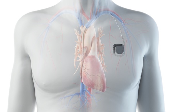
Leadless intracardiac pacemakers (LICPs) were developed to avoid some of the complications associated with traditional transvenous pacemakers, including:
- lead failure due to recurrent mechanical stress;
- injury to the tricuspid valve and/or subvalvular apparatus due to lead impingement; and
- infection of the leads or pacemaker pocket, which can result in bacteremia and endocarditis.1,2
In 2016, the FDA approved the Micra transcatheter pacing system (Medtronic) as an LICP to be implanted in the right ventricle (RV). The main complications associated with LICP placement include:
- RV rupture and pericardial tamponade;
- device dislodgment and embolization chiefly to the pulmonary artery, the right atrium or the superior vena cava; and
- complications related to vascular access.2
We report the rare occurrence of unintentional placement of a leadless pacemaker in the left side of the heart in a patient with a patent foramen ovale (PFO).
Case Presentation
The patient was a 67-year-old woman who presented for pacemaker insertion with a history of paroxysmal atrial fibrillation with rapid ventricular response and tachycardia-bradycardia syndrome with pauses up to 4.5 to 5 seconds. Her medical history was significant for metastatic left lung adenocarcinoma, and she had undergone left lung resection and radiation therapy to the left chest. She had suffered previous episodes of infection and bacteremia secondary to her immunocompromised state.
Since the patient had an infusion port in place with the catheter in the right internal jugular vein, the electrophysiology team believed that either infraclavicular approach would be suboptimal. They elected to place a leadless single-chamber permanent pacemaker via a femoral vein. During initial attempted placement along the RV mid-septum, sustained ventricular ectopy occurred and the device was not deployed there. On the second attempt, the thresholds were too high so the device was recaptured. On the third attempt, the device appeared to be in the appropriate position in the RV per fluoroscopic views in the right and left anterior oblique projections.
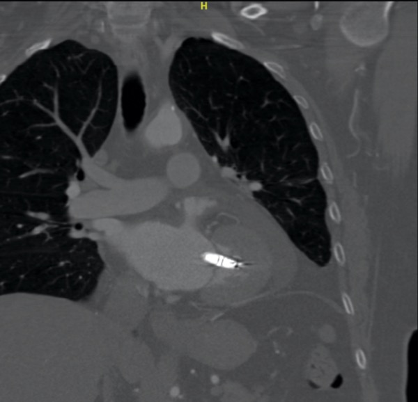
Five weeks later, the patient presented with progressive fatigue, shortness of breath and palpitations. Interrogation of the pacemaker showed no ventricular capture. Imaging with CT of the chest and PET as part of her cancer surveillance revealed the leadless pacemaker in the left ventricle (LV) (Figure 1). Subsequent transesophageal echocardiography (TEE) confirmed that the pacemaker was attached to the P1 and P2 cusps of the mitral valve extending into the LV with chordal involvement and moderate mitral regurgitation. A PFO with bidirectional flow and stable pericardial effusion were noted.
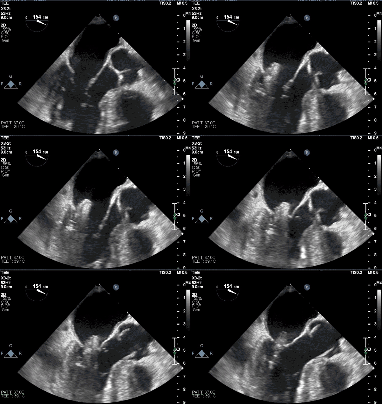
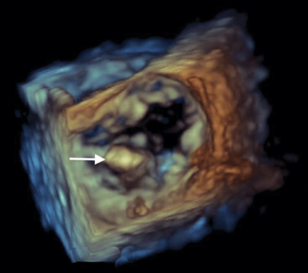
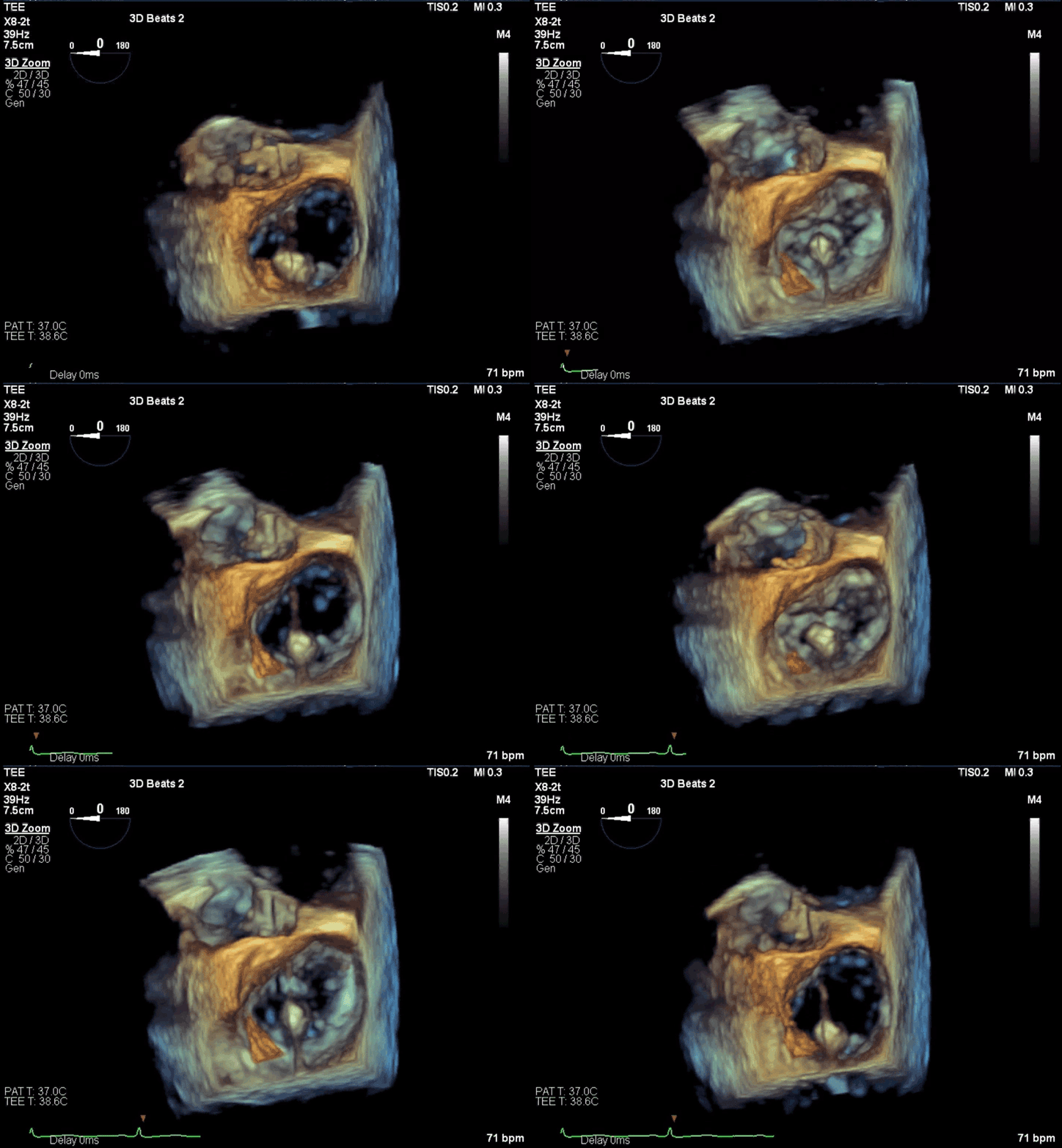
Since percutaneous retrieval of the pacemaker could confer a very high risk for further mitral valve damage, the decision was made to remove it surgically. The patient subsequently underwent removal of the LICP, mitral valve repair, PFO closure and epicardial pacemaker placement. Intraoperative TEE at the start of the case confirmed the attachment of the pacemaker to the P1 and P2 scallops of the mitral valve, and mild to moderate mitral regurgitation (Figure 2).
Discussion
Leadless pacemakers were originally approved for patients meeting the clinical criteria for RV pacing, but in 2020, the FDA approved the Micra AV pacemaker for use in patients with atrioventricular block.3 Today, LICP use is expanding to those in need of biventricular pacing.4 This increasing application of LICP technology emphasizes the need for better understanding of the complications associated with these devices.
The Micra Transcatheter Pacing Study Group reported a 1.6% incidence of cardiac perforation, 0.7% access-related issues, and 0.3% device dislodgment and venous thromboembolism.5 A 2021 case report by Ranka and colleagues describes an LICP that was found to be lodged in the lateral wall of the LV under the mitral valve after the patient presented with cerebellar infarcts consistent with a cardioembolic source; the authors concluded that the interatrial septum had likely been perforated at the time of insertion.6
To our knowledge, there are no other published cases reporting a leadless pacemaker intended for the RV but found in the LV in the setting of a PFO. In retrospect, our patient’s LICP was probably placed across the PFO at the time of initial insertion since multiple attempts were required and high thresholds were recorded. We concur with Ranka and colleagues who emphasize “the importance of analyzing orthogonal fluoroscopic views, contrast ventriculography, and paced electrocardiographic morphology during leadless pacemaker insertion to confirm accurate placement on the right ventricular septum.”6
The most likely reasons for a leadless RV pacemaker to be found in the LV include cardiac perforation during implantation5,6 or unintentional placement across an atrial or ventricular defect, as seen in our patient. The prevalence of a PFO in the general population is 25% to 30% and the majority are asymptomatic.7 Given the increasingly frequent application of LICP devices, it may be appropriate to consider whether there is a need to screen patients for PFO before they receive leadless pacemakers.
Conclusion
- Leadless pacemakers avoid some of the complications of traditional pacemakers and leads but may confer a greater risk for device malposition or embolization.
- Our case involving unintentional insertion of a leadless pacemaker in the LV via a PFO suggests that screening for PFO prior to device insertion may be helpful.
Sibert, a past president of the California Society of Anesthesiologists and member of the Anesthesiology News editorial advisory board, is the medical editor of “The Frost Series.” Authors who wish to submit a case to her may send it to FrostCaseSubmission@gmail.com. Please limit text to about 1,000 words and include an image, if at all possible.
References
- Hauser R, Gornick C, Abdelhadi R, et al. Major adverse clinical events associated with implantation of a leadless intracardiac pacemaker. Heart Rhythm Society. 2021;18:1132-1139.
- Lee J, Mulpuru S, Shen W. Leadless pacemaker: performance and complications. Trends Cardiovascular Med. 2018;28(2):130-141.
- Del Corral M, Covas P, Tracy C, et al. Percutaneous VDD leadless pacer implant post recent bioprosthetic tricuspid valve replacement for infective endocarditis. Pacing Clin Electrophysiol. 2021;44(4):747-750.
- Funasako M, Neuzil P, Duika L, et al. Successful implantation of a totally leadless biventricular pacing approach. HeartRhythm Case Rep. 2019;6(3):153-157.
- Reynolds D, Duray G, Omar R, et al. A leadless intracardiac transcatheter pacing system. N Engl J Med. 2016;374:533-541.
- Ranka S, Sheldon S, Downey P, et al. A case of leadless pacemaker in the left ventricle with cardioembolic stroke. JACC Clin Electrophysiol. 2021;7(4):563-564.
- Hagen P, Scholz D, Edwards W. Incidence and size of patent foramen ovale during the first 10 decades of life: an autopsy study of 965 normal hearts. Mayo Clin Proc. 1984;59(1):17-20.

