Weill Cornell Medical College - Qatar
Anesthesia, ICU and Perioperative Medicine Department
Hamad Medical Corporation
Clinical Anesthesiology Department
College of Medicine Qatar University
Doha, Qatar
Hamad Medical Corporation
Doha, Qatar
Hamad Medical Corporation
Doha, Qatar
Hamad Medical Corporation
Doha, Qatar
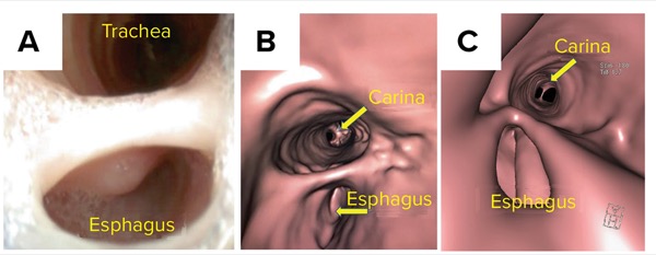
Hany A. Zaki, MD
Medical Emergency Department
Hamad Medical Corporation
Doha, Qatar
The complex anatomy of the airway has made proper assessment and evaluation a challenge, especially assessing distensibility and the potential collapse. The common pathway for respiratory and digestive systems is composed of potentially collapsible air-filled space, especially its proximal portion, with epithelial lined musculocutaneous components. This becomes of special concern in radiological assessment, and it is crucial to be fully aware of the detailed radiological anatomy—and the variants—for more accurate interpretation.
Virtual endoscopy (VE) of the airway has made remarkable progress in the past decade that has led to a significant clinical impact. The introduction of multidetector row CT scanners has made it possible to acquire high-resolution images of the upper, central and segmental airways within a short time, which can subsequently be reconstructed into elegant and distinguished 2D reformation and 3D images, including internal VE renderings that closely simulate images from conventional endoscopy (CE) or real endoscopy.
David Vining, MD, was the first to introduce and present the results of VE in 1994.1 Progress made with CT technology and the multidetector techniques allowed further applications to take place. A wide spectrum of applications is now established and clinically utilized including virtual colonoscopy, virtual bronchoscopy, virtual laryngoscopy, virtual otoscopy and virtual angioscopy.
Why VE?
Virtual endoscopy is a method to generate 2D and 3D images, similar to that obtained from CE examination, by reformatting the thin volumetric axial CT raw data set to be displayed on an electronic device using one of many commercially available types of VE software—either provided along with the CT scanners or even the open-source commercially available software, including RadiAnt, OsiriX or Horos.
This method has more advantages compared with CE. Virtual endoscopy is completely noninvasive with a better safety profile, and the resultant images are comparable with CE. There is no patient discomfort, no epistaxis, no gag reflex, no vomiting, and no potential aspirations or laryngeal edema and spasm. Potential hazards of CE include bleeding, pneumothorax, hypoxemia and aspiration; there are also risks associated with sedation when used. These risks opened the gates for new diagnostic risk-free tools, with VE being one.2 Many other potential hazards also may occur during CE. (3-5)
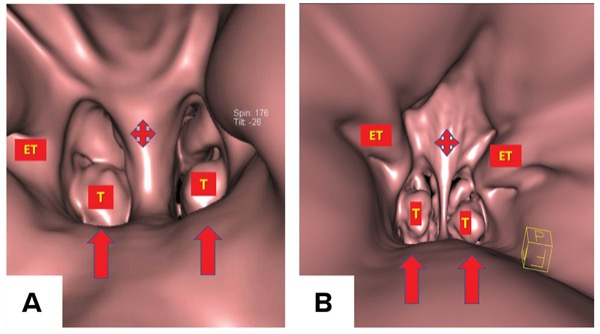
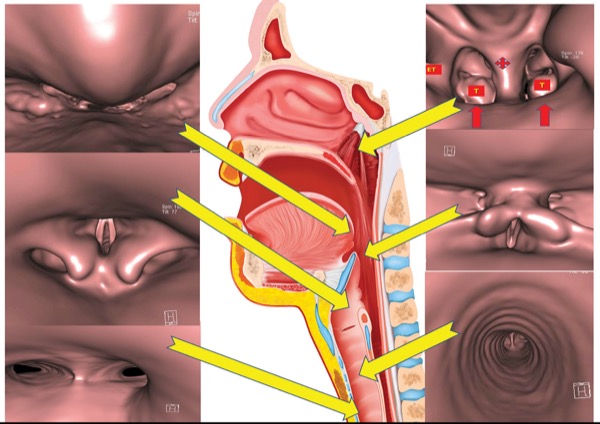
In addition to the aforementioned advantages over CE, VE has more advantages in getting perspectives that cannot be physically duplicated by any other means, for instance, visualization of the posterior choana, posterior nares and Eustachian tubes (Figure 1). It can also visualize the subglottic regions to assess for subglottic extensions of laryngeal neoplasm and can navigate and fly through in the caudocephalic direction. Virtual endoscopy can navigate and fly through narrow segments areas where CE cannot pass due to a relatively large caliber of the physical endoscope. Virtual endoscopy gives more valuable information that usually changes patient management.6
Limitations of VE
Despite all the advantages, VE has its flaws. It is still less sensitive to visualize any superficial lesion of the mucosal lining, which is inferior to CE.6 Virtual endoscopy cannot be used to assess mucosa, perform biopsies and therapeutic maneuvers. The presence of retained mucus or blood can falsely be reported as tracheobronchial stenosis or foreign body—a false overdiagnosis. Furthermore, the diameter of airway on the CT depends on the respiratory cycle (inspiratory/expiratory phases); therefore, stenosis of the tracheobronchial tree may be underestimated on inspiration. (It is crucial to perform VE during expiration to assess stenosis.) In some cases, such as tracheomalacia, assessment should take place during inspiration and expiration. Obtaining images during inspiration and expiration can be very challenging in examinations of infants and children.
Moreover, VE does not show the segmental and subsegmental parts of the tracheobronchial tree. It could be difficult or even impossible to detect some dynamic airway lesions, such as immobile vocal cords. There is a great debate regarding the time required to obtain the 3D reconstructed images and VE. It definitely has its learning curve, and with more practice that total time to obtain high quality would be around five to 10 minutes.6
Technique
The cornerstone of VE is the ability to obtain the best images without stairstep, respiratory motion, movement or coughing-related artifacts. The CT examination should include the area between the base of the skull down to below the carina level. Post-contrast series, with breath-hold and modified Valsalva maneuver, is essential in order to keep the potentially collapsible not-well-distended airway segments to be maximally distended. This will also alleviate the swallowing and coughing-related artifacts that might compromise the image quality.
Commercial VE software provides a navigation tool, “the virtual endoscope,” which dynamically navigates an organ lumen simulating the real scope. Virtual endoscopy allows the operator to “fly through” or “sail through” different directions and positions within the 3D anatomy of the sinonasal cavities. The virtual path is simultaneously displayed on a computer screen in axial, sagittal and coronal multiplanar reformation (MPR) images to make orientation easier.3,6
There are many techniques for generating the flight path during VE, including manual camera movement, semiautomatic path planning and automatic path planning. In manual camera movement, the position, field of view and focal point of the camera are interactively changed by the operator, with software providing collision detection to keep the camera within the potential cavity of the lumen. In semiautomatic path generation (also known as key-framing), the operator specifies key path points that have unique and directional coordinates and field of view, which are connected by cubic splines.
In the automatic flight path, at a rate of 25 to 30 frames per second, the computer generates an uninterrupted approximated flight route connecting the key views. The flight path is automatically computed by application after the operator designates an initial and one or more end points by computing the centerline of the imaged structure. The software registers the voxel coordinates of each key point in the specified shortest route to the goal end point and softens the path, avoiding obstacles including sinonasal, nasopharyngeal, oropharyngeal or laryngeal walls, which are recognized using predefined threshold ranges.
The typical flight path is planned through the air containing nostrils, nasal cavities, natural drainage pathways and ostia of paranasal sinuses, such as entering the maxillary sinus through the semilunar hiatus and frontal sinus through the frontal recess, and then navigating to the oropharynx down to the trachea, passing through different segments of the airway. While showing the partially transparent nasopharyngeal borders, the soft tissue could be made transparent to visualize the underlying bone morphology and preoperatively evaluate key structures like the carotid artery and optic nerve canals.
Manual flight generation might be needed in some cases, for example, in cases of mucosal swelling.3 Virtual endoscopy allows the operator to “journey” in different directions and positions within the 3D anatomy of the sinonasal cavities and visually demonstrated in axial, sagittal and coronal MPR images simultaneously to make orientation easier.6
With the camera panning around the nasopharynx, some views can be generated that are impossible by CE or mirror methods. Such views include—but are not limited to—the posterior aspect of nasal septum and turbinates, base of the tongue, uvula, tonsillar fossa, vallecula, pyriform sinuses and laryngeal structures including the vocal cords, trachea and carina, in a much clearer and more precise way.6,7
Evaluating the Normal Upper Airway
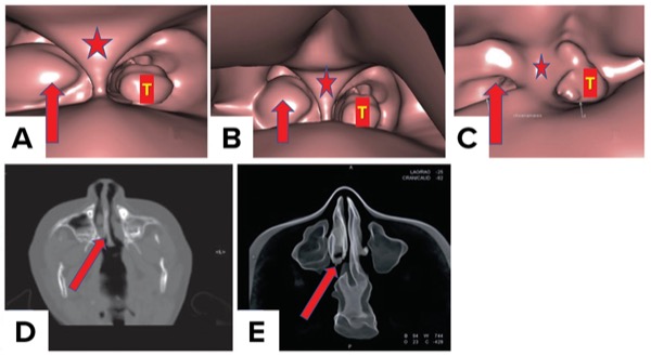
Virtual endoscopy evaluation through the anterior nasal orifice can show the columella, nasal orifices, nasal cavities, inferior and middle turbinates, meatus, osteomeatal complex orifice, nasal septum, posterior nares (choana), inferior turbinates, vomer, and Eustachian tube orifices. Diagnosis of the choanal atresia can be easily done using this technique within few seconds.
Virtual endoscopy of the normal nasopharynx from a posterior viewpoint provides a look at the Eustachian tube orifices in reference to the choanae and nasopharyngeal walls, an area that’s difficult to be seen with CE or any other diagnostic tool. Posterior choanal projection is an image that cannot be physically duplicated except by VE (Figure 1).
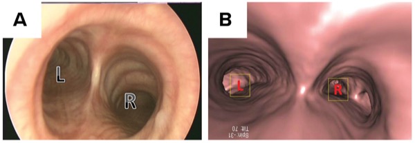
The trachea in an adult is usually 9 to 15 cm long and begins at approximately the sixth cervical vertebra (C6) at the inferior border of the cricoid cartilage (the only complete ring in the trachea). The inner diameter of the trachea is around 2.0 to 2.5 cm. The trachea is supported anteriorly by 16 to 20 C-shaped or incomplete cartilaginous rings. In the posterior view, the trachea consists of the pars membranacea, a flexible and mobile membrane that allows the trachea to change its configuration during both respiratory cycles (inspiration and expiration). Normally this membrane bulges during expiration, Valsalva maneuver and coughing. At the level of sternal angle (T4-T5), the main trachea divides at the level of the carina, a ridge formed by the downward and backward projection of the last tracheal ring, into the right and left main-stem bronchi (Figures 2 and 3).
During VE navigation, pyriform sinuses, aryepiglottic folds and vocal cords can be apricated, down to the trachea and carina. Radiologists, anesthesiologists and any other operator should be able to recognize the normal tracheal architecture and anatomic variants. In particular, normal structures such as the transverse aorta can indent or press the large airways and should not be confused with extrinsic lesions when viewed by CE.
Diagnosis of Congenital Nose Anomalies
A recognizable cause of posterior choanal obstruction is the congenital condition known as choanal atresia.
Choanal atresia can be a solitary finding or associated with other syndromes, such as CHARGE (coloboma, heart defects, growth retardation, genital hypoplasia and ear abnormalities). It represents with a range of symptoms, from acute obstruction and cyanosis to recurrent sinus disease. This depends on laterality and the presence of other congenital malformations (Figure 4).
Depending on the presentation, a number of investigations can be done to diagnose and assess the severity. Nasoendoscopy is preferred as an initial examination technique, as the point of obstruction can be identified and visualized. Thin-section CT is a great tool to assess the extent of obstruction and structures involved. In a recent study assessing the usefulness of helical 3D reconstruction and VE, Yunus et al observed that CT and VE are valuable for defining the type and extent of the disease, as VE of the nasopharynx from a posterior view can provide the view of Eustachian tube opening with reference to bony borders of the nasopharynx.8 Preoperative evaluation of the condition using CT and VE would be helpful in surgical planning.
Diagnosis of Pharyngeal Lesions
Virtual endoscopy and 3D reconstruction of the airway can be used to diagnose and stratify patients with obstructive sleep apnea to show the pharyngeal collapsibility without the need for invasive CE or nasoendoscopy.9 Another area in which VE can be useful is pharyngeal masses, especially when it is risky to perform an awake fiberoptic examination (e.g., high risk of bleeding). In such cases, VE can assist in planning the anesthetic management with minimal risk.10,11
The case reported below clearly shows the usefulness of VE in solving the mystery of a difficult airway case in our center. A 32-year-old man was referred to our difficult airway clinic (DAC), complaining of difficulty in swallowing and abnormal “pinprick” sensation in his throat for more than 18 months.
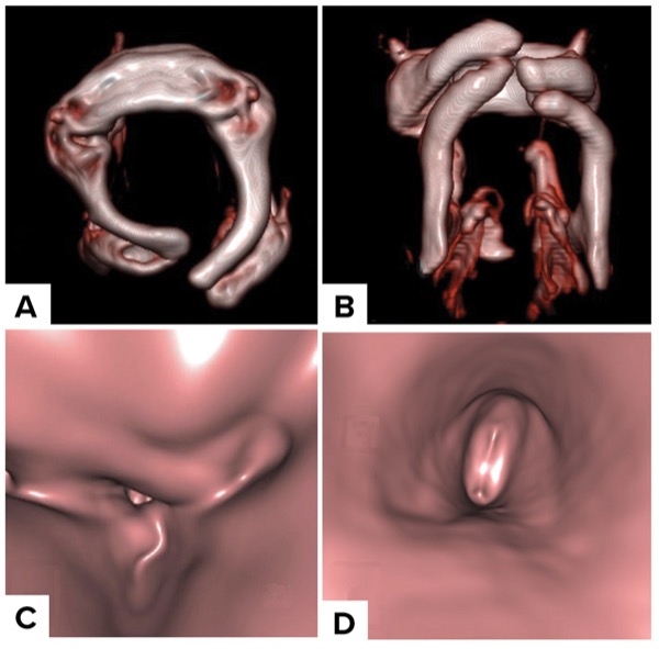
A nasoendoscopy evaluation showed prominent corniculate cartilages and a left vocal cord polyp. A CT scan of the larynx showed normal vocal cords and a thick abnormal hyoid bone causing narrowing and indenting on the posterior upper hypopharyngeal wall. A 3D reconstruction was done confirming the abnormal hyoid bone shape and narrowing of the pharyngeal area (Figure 5).12
Diagnosis of Epiglottic and Supraglottic Lesions
Beside the principal diagnostic tools in the evaluation of the supraglottic diseases, VE can be extremely useful in diagnosis and extent assessment with minimal risk to patients. It does not require extra investigations since it uses the already present CT images.13 The two patients we present below illustrate this clearly.
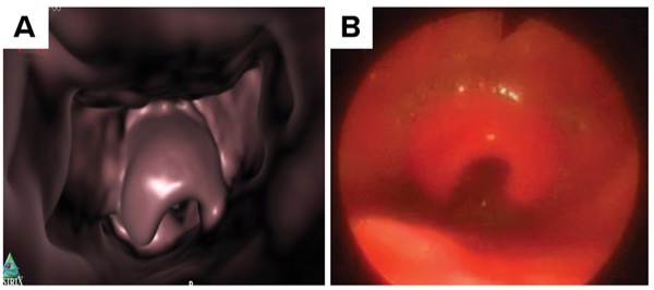
Figure 6 shows identical views by CE and VE for a patient with epiglottitis. Although the VE images lack the red angry look of CE, it still can provide a very informative image, with much safer approach in such a situation with a high risk for airway obstruction.
Diagnosis of Supraglottic Cyst
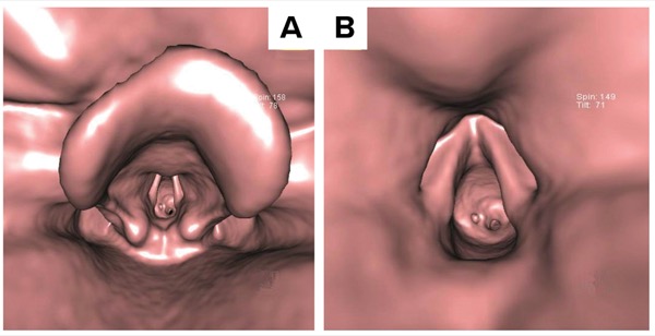
Virtual endoscopy can be extremely helpful in supraglottic masses diagnosis and extent assessment (Figures 7). There is the normal appearance of the vallecula and epiglottis. However, there is encroachment on the supraglottic space by a mass lesion appreciably reducing its caliber. Further navigation beyond the mass shows normal caliber of the trachea.
Diagnosis of Supraglottic Web or Membrane
Virtual endoscopy can successfully detect laryngeal lesions, with good image quality.11 It is an additive tool in conjunction with direct laryngoscopy—when feasible—and easily reaches the lower limit of the lesion compared with only direct laryngoscopy.14 Virtual endoscopy is also sensitive in assessment of the larynx beyond large supraglottic and glottic masses obscuring the view of CE.15
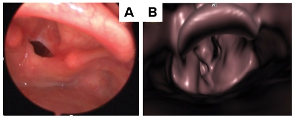
Note the identical views of a vocal cord web (post-traumatic) in both CE and VE (Figure 8). This demonstrates how handy VE can be if the patient refuses CE. It also shows how sensitive VE can be in detecting laryngeal lesions. We also note another good example of one case of traumatic supraglottic stenosis easily diagnosed with VE compared with CE (Figure 8).16
Uses in Glottic and Vocal Cord Lesion
Virtual endoscopy is a very useful adjunctive radiological tool in the assessment of laryngeal lesions, as it does not require general anesthesia, allows 3D visualization of the airway beyond areas of narrowing and gives a highly accurate representation of vocal cord lesions—both in terms of definition and spatial representation.17 Virtual endoscopy can also direct biopsy planning of cord lesions, and the 3D location of polypoid lesions is easier to appreciate.17 VE is very useful in the diagnosis of epiglottitis without doing Conventional endoscopy and complicate the clinical scenario (Figure 6).
Uses in Subglottic Stenosis
Subglottic stenosis can result from an endoluminal lesion or extraluminal compression. The differentiation between both can be difficult—or even misleading—when using CE. Virtual endoscopy provides a useful tool to evaluate the stenotic lesion and its relation (Figure 9).18
Abo Elnaga et al found the sensitivity, specificity and diagnostic accuracy of virtual bronchoscopy in localizing the site of stenosis were 100% for all. The sensitivity, specificity and diagnostic accuracy of virtual bronchoscopy in grading the stenosis were 85.3%, 94.5% and 92%, respectively. The sensitivity, specificity and diagnostic accuracy of virtual bronchoscopy in measuring the length of stenosis were 92.3%, 94.3% and 92%, respectively.19 However, the researchers concluded that confirmation is needed by rigid bronchoscopy for measurement of the grade and length of stenosis.
Uses in Tracheal Lesions
Conventional endoscopy visualizes the mucosal layer and is an important diagnostic and therapeutic tool. However, assessing the deep layers or the adjacent tissue is not possible. Virtual endoscopy provides an excellent evaluation of the airway wall.20 Moreover, multidetector CT is now considered a first-line screening tool for clinically suspected tracheobronchomalacia and may also serve as an adjunct to bronchoscopy in preoperative planning or even as an alternative to bronchoscopy in the pediatric or older populations.21 The acquisition technique is important to maximize the ability of the study in the diagnosis and classification of patients. At end expiration, tracheal collapse is submaximal; therefore, dynamic acquisition during expiration must be used whenever the patient can follow instructions.22
Airway Foreign Body Management
Airway foreign bodies (AFBs) is an interdisciplinary area between emergency medicine, pediatrics and otolaryngology. It is a life-threatening condition that is not uncommon, as accidental aspiration of an element into airways is a widespread clinical scenario among children younger than 3 years of age, is the leading cause of infantile deaths and the fourth leading cause of death among preschool children.23,24
The incidence is lower in adults than in pediatric patients. A CT scan can provide information regarding the location and characteristics of FBs, and aid in diagnosis, yet bronchoscopy remains the gold standard for diagnosis and management of FB aspiration.25
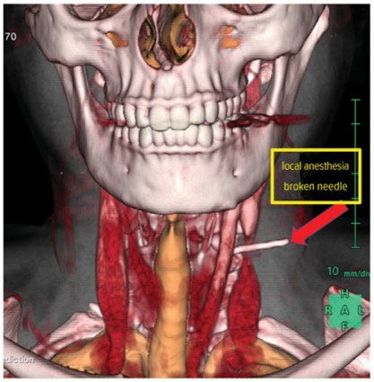
Virtual endoscopy can contribute to the diagnosis of AFBs and management planning, as in a case that presented to our center. A middle-aged female patient complained of throat pain that had lasted a couple of weeks. She claimed that it all started immediately after having duck for lunch, assuming she had swallowed a duck bone.
A thorough physical examination was performed, including CE, but did not show any evidence of an FB in the upper aerodigestive tract. There was a tender, non-fluctuant, diffuse swelling of about 2 by 2 cm on the left side of the neck. An x-ray was taken and revealed a long, thin, smooth and impinged FB in the neck, lying lateral to the esophagus—not closely resembling a duck bone.
Of note, 3D reconstruction, VE images and video were produced from a CT scan and very useful for detecting the FB (Figure 10). It was a broken dental anesthesia needle, as the patient had a dental procedure around the same time of pain but did not connect the pain to the procedure.26
With VE, we noticed proximally the vallecula, epiglottis and pyriform sinuses, then navigation through both true and false vocal cords, which were of average normal anatomy. Then further distally, we saw the caliber of the trachea, which was within average normal showing mild indentation by the tip of the needle on the left side.
Tracheal narrowing (either congenital or acquired) may result from intrinsic tracheal stenosis and/or external compression, at any anatomic level. Alteration in the tracheal diameter changes the airflow dynamics into turbulent flow. Tracheal compression can be caused by several factors, for example major vascular anomalies, bronchogenic cyst, an enteric duplication cyst, thoracic neurogenic tumor and/or a mediastinal teratoma.
Chest radiograph, barium swallow examination, bronchoscopy, CT and MRI are several methods of diagnosis.27
Uses in Tracheal Stenosis Diagnosis in COVID-19 And Other Causes
Tracheal stenosis often results in varying degrees of distortion along the vertical plane in addition to airway narrowing, which predisposes to airflow turbulence.28
The COVID-19 pandemic created a huge volume of patients with prolonged intubation history, resulting in a surge in cases of tracheal stenosis.29
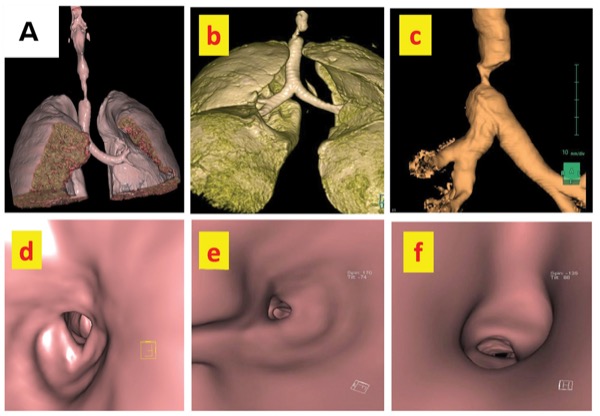
Figure 11 shows 3D and VE images for one of our post–COVID-19 tracheal stenosis cases. It starts at the level of the vallecula, epiglottis and upper airway, then passing through normal-caliber vocal cords and proximal trachea followed by abrupt reduction of the caliber of the trachea due to stenosis. Sometimes it is quite difficult to pass through a narrow lumen using CE, which is not the case in VE, where we can pass through millimeter-wide narrowing. Shortly after, the trachea gained its normal caliber down to the carina and bifurcation. With the advent of VE, we can reverse the direction of the endoscope upward toward the base of the tongue, nasopharyngeal airway, posterior nares, inferior nasal turbinates and nasal septum.
Other conditions such as tumors (both benign and malignant) and various autoimmune and inflammatory diseases can cause tracheal stenosis in adult patients. Because of the wide adoption of low-pressure cuff endotracheal tubes, the total incidence of stenosis has fallen to less than 1% (in the pre–COVID-19 practices). High cuff pressures lead to tracheal mucosal necrosis, followed by scarring and eventually stenosis. Tracheal stenosis following tracheostomy or prolonged intubation frequently occurs at two locations: at the level of the cuff of the endotracheal tube and at the stoma site (Figure 11).27
Diagnosis of Tracheoesophageal Fistula
Tracheoesophageal fistula (TEF) is an abnormal communication between the trachea and the esophagus. Causes of TEF are numerous and could be classified into congenital and acquired. Patients typically present with cough after deglutition and/or recurrent chest infections/aspiration pneumonia. The latter is a reason for increased morbidity and mortality of TEF. Therefore, early detection and appropriate management are vital.
Esophagoscopy and bronchoscopy are the gold standard for diagnosis. Radiological studies using VE and 3D reconstruction are important to plan surgical intervention. Size, location and depth of the fistula are important factors to consider when planning the best approach for surgicalclosure or to plan the use of regional flaps (Figure 12).30
Conclusion
Virtual endoscopy is a technique for creating computer simulations of anatomy from radiological image data and viewing those simulations in a way that is similar to CE and helpful to orient the image to correspond with a conventional endoscopic view.
Virtual endoscopy is a powerful tool in diagnosis of airway-related pathologies and planning of treatment strategies, and it has its place in the recent practice. It still has its own drawbacks, but this can be easily overcome with development and evolution of technology.
The uses of VE are not limited to diagnostics. A combination of VE and CE in one field using Microsoft HoloLens, in which the image will be displayed in a 3D hologram mode, with the possibility of rotating and moving it at will at any time, can be a viable tool for teaching and training on CE.29
The evolution and advancement in both radiological image acquisition and software post-processing, through machine learning and artificial intelligence, will definitely improve the technology of VE and image quality, and overcome the flaws in it. This might not eliminate the need for CE entirely, especially for therapeutic and biopsy indications, but will definitely decrease the need for diagnostic-only CE.
References
- AJR Am J Roentgenol. 1994;162(suppl):104.
- Radiology. 1996;200(1):30-31.
- Free-flight: a virtual endoscopy system. In: Lecture Notes in Computer Science. Vol 1205. Springer; 1997.
- J Clin Diagn Res. 2015;9(4):TC14-TC18.
- Role of virtual endoscopy and 3-D reconstruction in airway assessment of critically ill patients. In: Virtual Endoscopy and 3D Reconstruction in the Airways. Springer; 2023.
- Radiological evaluation of the airway: one-stop shop. In: Virtual Endoscopy and 3D Reconstruction in the Airways. Springer; 2019.
- J Comput Assist Tomogr. 2004;28(5):661-669.
- J Pak Med Assoc. 2012;62(11):1154-1160.
- J Kanazawa Med Univ. 2004;29:243-251
- Br J Anaesth. 2014;113(2):304-306.
- J Korean Radiol Soc. 2000;42(5):715-720.
- Trends Anaesth Crit Care. 2017;16:27-28.
- J Digit Imaging. 2007;20(1):67-71.
- Egyptian J Radiol Nuclear Med. 2013;44(3):497-503.
- Mohammed, H., Haidar, H., Shallik, N.A., Elhakeem, A., Al Abdulla, M., Dogan, Z. (2019). Virtual Endoscopy and 3-D Reconstruction/Prototyping in Head and Neck Surgeries. In: Shallik, N.A., Moustafa, A.H., Marcus, M.A.E. (eds) Virtual Endoscopy and 3D Reconstruction in the Airways. Springer, Cham. https://doi.org/ 10.1007/ 978-3-030-23253-5_8; 2019
- Trends Anaesth Crit Care. 2017;16:16-17.
- Clin Otolaryngol Allied Sci. 2002;27(2):98-100.
- Postgrad Med J. 2000;76(893):187-188.
- J Otolaryngol ENT Res. 2016;5(2).
- Insights Imaging. 2017;8(2):255-270.
- Ann Diagn Pathol. 1997;1(1):31-46.
- Pediatr Radiol. 2008;38(1):82-88.
- World J Emerg Med. 2016;7(1):5-12.
- J Otolaryngol Head Neck Surg. 2008;138:576-580.
- J Thorac Dis. 2017;9(9):3398-3409.
- J Dent Anesth Pain Med. 2020;20(2):83-87.
- Surgical disorders of the chest and airways. In: Avery’s Diseases of the Newborn. 9th ed. W.B. Saunders; 2012.
- J Thorac Dis. 2014;6(3):258-270.
- PLoS One. 2014;9(2):e89224.
- Challenging clinical cases discussion. In: Virtual Endoscopy and 3D Reconstruction in the Airways. Springer; 2019.
Copyright © 2024 McMahon Publishing, 545 West 45th Street, New York, NY 10036. Printed in the USA. All rights reserved, including the right of reproduction, in whole or in part, in any form.
Download to read this article in PDF document:![]() Virtual Endoscopy, a Powerful Tool for Airway Pathology and Treatment Strategies
Virtual Endoscopy, a Powerful Tool for Airway Pathology and Treatment Strategies



Please log in to post a comment